Last Updated on August 23, 2025 by Max
Introduction
The prostate is a small gland with a big impact. Weighing only 15–20 grams in young men—about the size of three almonds—it sits directly below the bladder and wraps around the urethra. Despite its modest size, it influences both urination and fertility. By their 60s, almost half of men experience urinary changes linked to prostate enlargement (Barry, M.J., 2020).
Understanding how the prostate interacts with surrounding structures makes sense of these changes. On this page, you’ll find an interactive diagram you can explore, along with a detailed guide explaining the anatomy. Together, they show why this walnut-sized gland plays such a central role in male health.
Explore the Prostate: Interactive Diagram
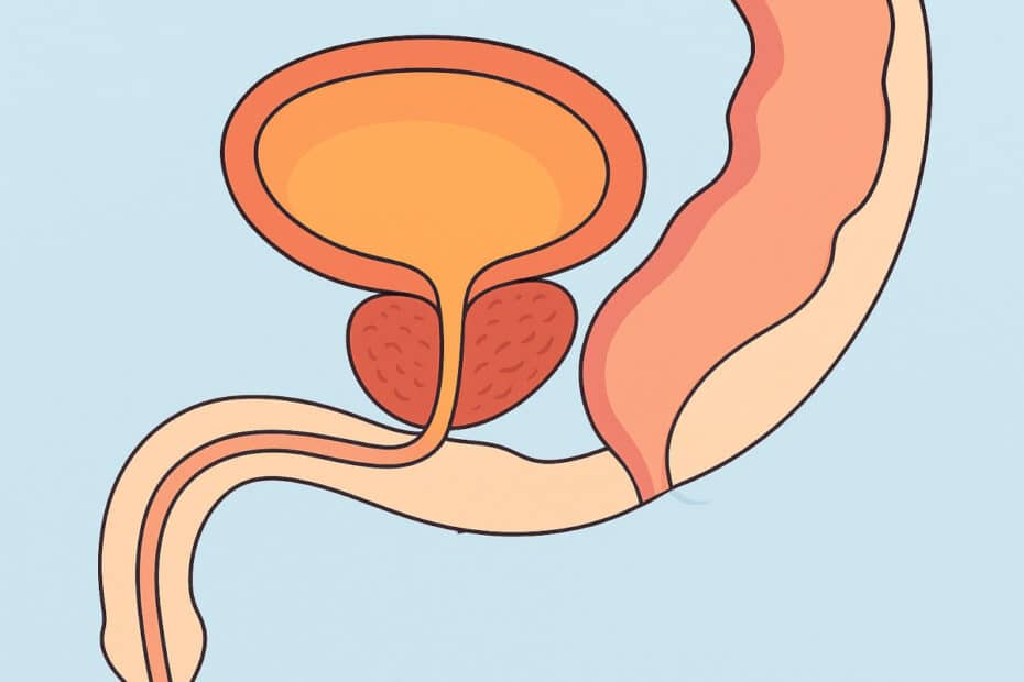
Use the interactive diagram below to tap or hover over each part—the prostate, bladder, urethra, seminal vesicles, rectum, and surrounding nerves. Each label highlights what the structure does and how it relates to prostate health.
When you click a label, you’ll jump to the corresponding explanation below. This way, you can explore visually first, then read deeper details for context.
Explore the Prostate: Interactive Diagram
Tap or hover to learn. Click a label to open details, then jump to the full explanation below.
Prostate Gland
The prostate begins as a walnut-sized gland weighing about 15–20 grams. With age, it often enlarges, sometimes exceeding 100 grams in advanced benign prostatic hyperplasia (BPH) (Barry, M.J., 2020). Located just beneath the bladder and wrapped around the urethra, it produces about 30% of semen. Its secretions are slightly alkaline, protecting sperm in the acidic vaginal environment. Surprisingly, sperm themselves account for less than 5% of semen; the rest comes from the prostate and seminal vesicles (Mann, T., 2020).
As the prostate grows, it compresses the urethra. Even a few extra grams of tissue narrow the passage enough to slow urine flow, explaining symptoms such as weak stream, hesitancy, or dribbling.
Bladder
The bladder is a muscular reservoir that stores urine, typically signaling fullness at 300 ml but capable of stretching close to 600 ml. Positioned directly above the prostate, its outlet passes through the gland. This setup means even modest prostate growth can disrupt normal emptying. Over time, the bladder muscle thickens and strains to push urine past the narrowed outlet. That stress often results in nocturia—waking up at night to urinate—which affects nearly 70% of men over 60 (Irwin, D.E., 2020).
Urethra
The urethra functions like a straw running straight through the prostate. Any gland enlargement squeezes this channel interfering with urine flow. Clinically, flow rates fall noticeably with only minor narrowing, which physicians measure using uroflowmetry (Gravas, S., 2022). The urethra also serves as the final pathway for semen, making it a shared channel for both urinary and reproductive systems.
Seminal Vesicles
The seminal vesicles, each about 5 cm long, sit behind the bladder and above the prostate. They contribute roughly 60% of semen volume, producing fluid rich in fructose—the primary energy source for sperm. Without this sugar, sperm lose motility and their ability to reach the egg (Mann, T., 2020). Prostaglandins in the vesicular fluid thin cervical mucus, further supporting fertilization. These glands’ close relationship with the prostate means inflammation or blockage often affects ejaculation.
Vas Deferens and Ejaculatory Ducts
The vas deferens is a muscular tube that propels sperm from the testes at speeds near 5 meters per hour during ejaculation (Carson, C.C., 2021). As it nears the prostate, it merges with the duct of the seminal vesicle to form the ejaculatory duct. These ducts pierce the prostate and empty into the urethra, mixing sperm with prostate and seminal vesicle secretions. Enlargement or swelling of the prostate can compress these ducts, altering semen volume and sometimes causing painful ejaculation.
Rectum
Directly behind the prostate lies the rectum, separated by only a thin tissue wall. This proximity allows physicians to assess the prostate with a digital rectal exam (DRE). Despite its simplicity, the DRE remains valuable; studies show it detects abnormalities in up to 18% of cases missed by PSA alone (Cornu, J.N., 2021). The rectum’s alignment with the gland also makes it a key access route for imaging and biopsies.
Nerves and Blood Supply
The prostate is enveloped by delicate networks of blood vessels and nerves. Among them are the cavernous nerves, critical for erectile function. Because these bundles run along the prostate’s capsule, surgery in this area must be extremely precise. Damage explains why erectile dysfunction sometimes follows prostate surgery. Advances like nerve-sparing prostatectomy protect these bundles, with studies showing potency preserved in more than 70% of properly selected patients (Walz, J., 2020).
Why This Anatomy Matters. When you look at these structures together, the story becomes clear. The prostate’s location—beneath the bladder, around the urethra, and next to the seminal vesicles and rectum—makes it a central player in both urinary and reproductive health. As it grows, it not only disrupts urination but also alters semen flow, bladder function, and sometimes sexual performance. Recognizing this interplay explains why prostate disorders have such wide-ranging effects on quality of life.
References
- Barry, M.J., 2020. Epidemiology and natural history of benign prostatic hyperplasia. Urology Journal.
- Mann, T., 2020. Biochemistry of Semen and the Male Reproductive Tract. Cambridge University Press.
- Irwin, D.E., 2020. Prevalence of nocturia and its impact. BJU International.
- Gravas, S., 2022. Guidelines on the management of non-neurogenic male lower urinary tract symptoms. European Association of Urology.
- Carson, C.C., 2021. Pathophysiology of Ejaculatory Disorders. Andrology Journal.
- Cornu, J.N., 2021. Evaluation of the Prostate via Digital Rectal Examination. Urology Clinics.
- Walz, J., 2020. Nerve-sparing techniques in radical prostatectomy: Evolution and outcomes. European Urology.
I have had a Tulsa Pro procedure (MRI guided ultrasound ablation) for prostate cancer 18 months ago. I have a neurogenic bladder requiring self- catheterizations 4-5 x per day.
My urologist can not pass a cystoscope into my bladder. He wants to do a TURP.
8 months ago I developed a false passage and changed my straight catheter to a crude catheter and can pass the crude catheter to my bladder without a problem to pass my urine. There is no hematuria.
Question: Will a prostate MRI be able to show my prostatic urethra that is patent or obstructed?
Is it possible that the cystoscope was in my false passage and not in my prostatic urethra?
Again, I can pass my crude catheter into my bladder without problems.
THx, Bill Morgan, MD
Hi Bill,
Thanks for your question and for explaining your situation in detail. I’m not a urologist or surgeon, but I do research and write about prostate health, so I can share what current evidence and clinical experience suggest.
Regarding your first question: a prostate MRI generally isn’t very good at showing whether the prostatic urethra is truly open or blocked. MRI is excellent for viewing the prostate and any changes from treatments like TULSA-Pro, but the urethral channel is very small, and MRI usually can’t show small strictures, false passages, or whether a scope can physically pass through. It may show major scarring or distortion, but it can’t reliably confirm urethral patency. When doctors need a clear picture of the urethra, they usually rely on studies like a retrograde urethrogram or a voiding cystourethrogram, or they repeat cystoscopy under better guidance.
As for whether your cystoscope might have gone into the false passage: yes, that’s very possible. When someone has a known false tract from catheterization, a rigid cystoscope can be accidentally steered into it—especially if there is any narrowing or scarring in the true urethra after treatments like TULSA. What you describe actually makes sense. The fact that you can easily pass a coudé catheter, without bleeding, strongly suggests that your real urethral channel is still open. The curved tip of a coudé catheter tends to follow the natural pathway of the urethra and is much less likely to slide into a false passage.
People often wonder why a catheter can pass but a cystoscope cannot, and the explanation is simple. A catheter is smaller, more flexible, and easier to guide. A cystoscope is larger, rigid, and much easier to misdirect into a false passage. Even a small ring of scar tissue from healing after TULSA-Pro can stop a scope, while still allowing a catheter to slide through.
Because of all this, it isn’t a given that TURP is the right next step. A TURP helps when there is prostate tissue causing blockage, but it doesn’t fix a false passage or a urethral stricture. That’s why, before going ahead with surgery, most urologists would want clearer imaging of the urethra—usually through a RUG or VCUG—or a repeat cystoscopy under anesthesia or with guidewire assistance. This helps determine whether the problem is prostate tissue, scar tissue, or simply the scope being redirected.
In short, MRI won’t give a definitive answer about the urethra. It is entirely possible that the cystoscope went into the false passage. And your ability to pass a coudé catheter smoothly is a good sign. Before deciding on TURP, it would be reasonable to have the anatomy clarified more precisely.
Hope this helps, and I wish you the best as you sort it out with your urologist.